Chronic obstructive pulmonary disease: exercise is not necessarily beneficial
Release date: 2015-02-26 Chronic obstructive pulmonary disease (COPD) is a complex condition with chronic cough. The patient's fistula can reflect airway inflammation. More importantly, multiple alveolar damage, limited airflow, and subsequent diagnosis of systemic inflammation and Extrapulmonary reaction such as muscle atrophy. Inflammation of COPD involves a number of different cells and molecules, the extent and severity of which can be estimated by measuring the concentration of oxidative stress and inflammatory mediators, acute phase proteins. For example, C-reactive protein (CRP), an acute phase protein synthesized primarily by hepatocytes, is a marker of poor lung function caused by tissue damage and inflammation, leading to mortality in COPD patients. Tumor necrosis factor alpha (TNF-alpha) plays an important role as a marker of cancer, chronic heart failure and cystic fibrosis. Scholars such as Amani I. El Gammal in Ireland, Switzerland and Canada have three goals in this study: first, to confirm the relationship between large-scale, systemic biomarkers and the severity of COPD; second, to examine sharply The effects of exercise on these biomarkers; third, examining the effects of pulmonary rehabilitation on these biomarkers. The researchers measured high-sensitivity CRP, TNF-α, interleukin-6 (IL-6), transforming growth factor beta (TGF-β), WCC, neutrophils, and oxidative stress. Researchers have published relevant research in the English Journal of Open Journal of Respiratory Diseases. The study population consisted of 40 patients and excluded patients with the above conditions. The study began on August 1, 2006 and ended on June 30, 2008. During the study, the researchers performed lung function and cardiopulmonary exercise tests on these 40 people, and also carried out a lung rehabilitation program to measure their health, including cytokines and oxidative stress. Inflammatory response after rapid exercise in patients with COPD After exercise, members of the COPD group were an important factor in neutrophil counts compared with healthy controls, with leukocytosis. However, they themselves have more white blood cells than healthy subjects, so the results of COPD patients are not exaggerated. Unlike the control group, if the systemic inflammatory response is elevated in patients with COPD, the WCC value does not return to baseline after exercise. The IL-6 value of the COPD group increased after exercise, while the healthy control group did not have a similar change. The serum concentration of TGF-β1 increases with the severity of the condition, confirming its role as a marker of the onset of the disease. However, the link between TGF-β1 and structural changes in COPD is not obvious, and there may be more complicated relationships. Pulmonary rehabilitation treatment effect Cytokine levels did not change after eight weeks of rehabilitation. The trained lateral femoral muscle maintained a higher glutathione level (GSH, the most important antioxidant) compared to the untrained state, so the training appeared to increase the antioxidant buffering effect of the control group. However, the COPD group did not receive these benefits, which means that the patient group could not achieve the same effect as a healthy person. To make matters worse, training increases the level of oxidized GSH (GSSG). Measurements were made on the trained COPD group, and the condition of inflammation increased during and after intensive exercise. The type, density and duration of exercise are highly correlated with differences in cytokines. Therefore, scholars have concluded that patients with chronic obstructive pulmonary disease have higher levels of systemic inflammation, and markers of inflammation are associated with the severity of the disease. Exercise responded more strongly to inflammation in the COPD group, but it was not so exaggerated. Scholars hypothesized that pulmonary rehabilitation changes inflammatory mediators, as long-term training is healthy, and this study cannot be confirmed. Source: Thousand people think tank
Human chorionic gonadotropin is a hormone for the maternal recognition of pregnancy produced by trophoblast cells
Human chorionic gonadotropin (HCG) is a glycoprotein secreted by trophoblast cells of the placenta And β dimer proteins. The molecular weight of glycoprotein hormone 36700, A. Pituitary, FSH, follicle stimulating hormone, LH (luteinizing hormone) and TSH (thyroid stimulating hormone) are basically similar, So they can cross-react with each other, while the structure of β subunit is not similar. In mature women, the fertilized ova move to the uterine cavity for implantation and form embryos. In the process of development and growth into the fetus, the placental syncytiotrophoblast cells produce a large amount of HCG, which can be excreted into urine through the blood circulation of pregnant women. Serum and urine HCG levels rise Rapidly from 1 to 2.5 weeks of gestation, reaching a peak at 8 weeks of gestation and dropping to moderate levels at 4 months of gestation, which remain at the end of gestation. At present, the commonly used detection methods are: latex aggregation inhibition test and hemagglutination inhibition test; Radioimmunoassay (RIA); Enzyme linked immunosorbent assay (ELISA); Monoclonal antibody colloidal gold test
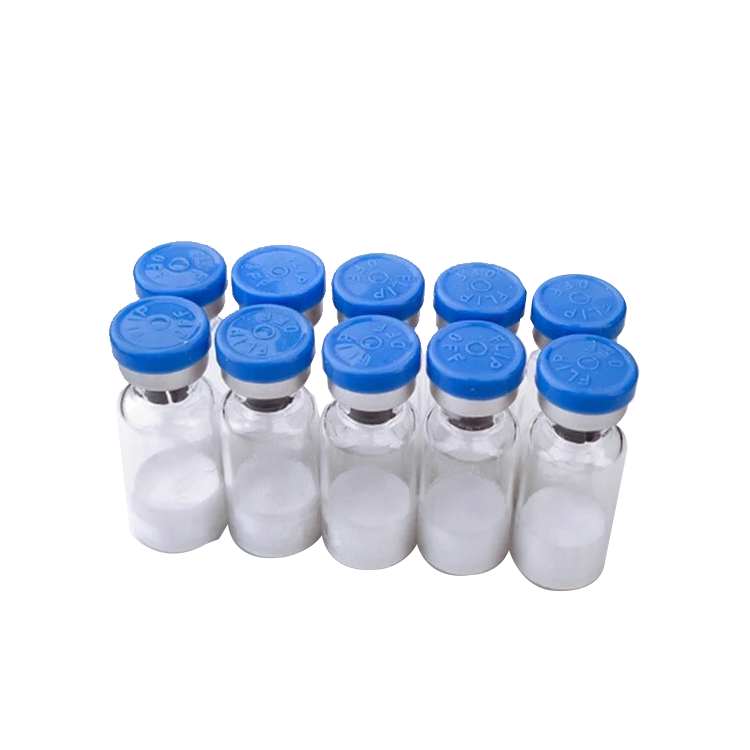
Hcg Powder,Custom Ghrp Peptides,High Purity Hcg Powder,Popular Peptide Bodybuilding Powder Shaanxi YXchuang Biotechnology Co., Ltd , https://www.peptide-nootropics.com
