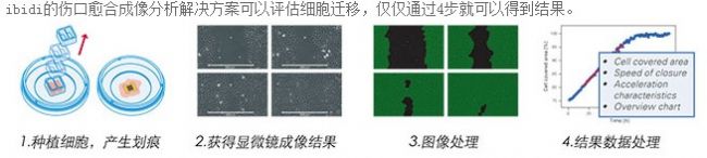
A brief introduction to a new experimental method of cell scratch
colostomy pouch ostomy pouch stoma pouch urostomy pouch Wenzhou Celecare Medical Instruments Co.,Ltd , https://www.celecaremed.com
1. Basic principles
The cell scratch (repair) method is a simple method for determining the migration and repair ability of cell migration, similar to the in vitro wound healing model, in a single cultured dish or plated monolayer adherent cells, using a micro-tip or other hard object in the cell. The central region of the growth is streaked, the cells in the central part are removed, and then the cells are cultured for the time set by the experiment (for example, 72 h), and the cell culture plate is taken out to observe whether the surrounding cells grow (repair) to the central scratch zone, thereby judging The ability of cells to grow and migrate, the experiment usually needs to set the normal control group and the experimental group. The experimental group is a group with some treatment factors or drugs, exogenous genes, etc., through the cells between different groups for the scratched area. The ability to repair can determine the ability of each group of cells to migrate and repair. 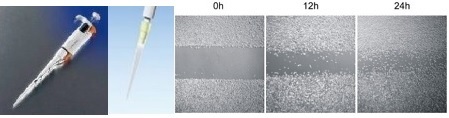
Experimental Materials Cell sample Reagents, kits Serum-free medium PBS Instruments, consumables 6- hole board maker pen  ruler  Gun head Experimental procedure
1. First use the marker pen on the back of the 6-hole plate, with a ruler to evenly draw a horizontal line, about every 0.5-1cm, across the hole. Pass at least 5 lines per hole.
All sterilizable instruments should be sterilized, and the ruler and marker pen should be irradiated with ultraviolet light for 30 minutes before operation.
2. Add about 5X10 5 cells in the air, the specific number varies from cell to cell, and it can be covered by overnight.
3. The next day, use the tip of the gun to measure the horizontal line as far as possible. The tip of the gun should be vertical and not tilted.
4. Wash the cells three times with PBS, remove the cells, and add serum-free medium.
5. Place in a 37 degree 5% CO 2 incubator and incubate. Take samples at 0, 6, 12, 24 hours and take pictures.
2 , the operation steps
(1) First use the marker pen on the back of the 6-hole plate, with a ruler, evenly draw the horizontal line, about every 0.5~1cm, across the hole. Pass at least 5 lines per hole.
(2) About 5 × 10⁵ cells were added to the well, and the specific amount was different depending on the cell type. The principle of inoculation was that the fusion rate reached 100% after overnight.
(3) The next day, use the tip of the gun to measure the horizontal line as far as possible. The tip of the gun should be vertical and not tilted (it is best to use the same gun head between different holes).
(4) The cells were washed 3 times with PBS, and the cells were removed, and serum-free medium was added.
(5) Place in a 37 ° C 5% CO2 incubator and incubate. You can take samples at 0, 6, 12, 24h time points and take photos (the specific time depends on the needs of the experiment).
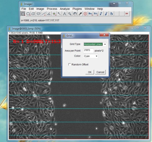
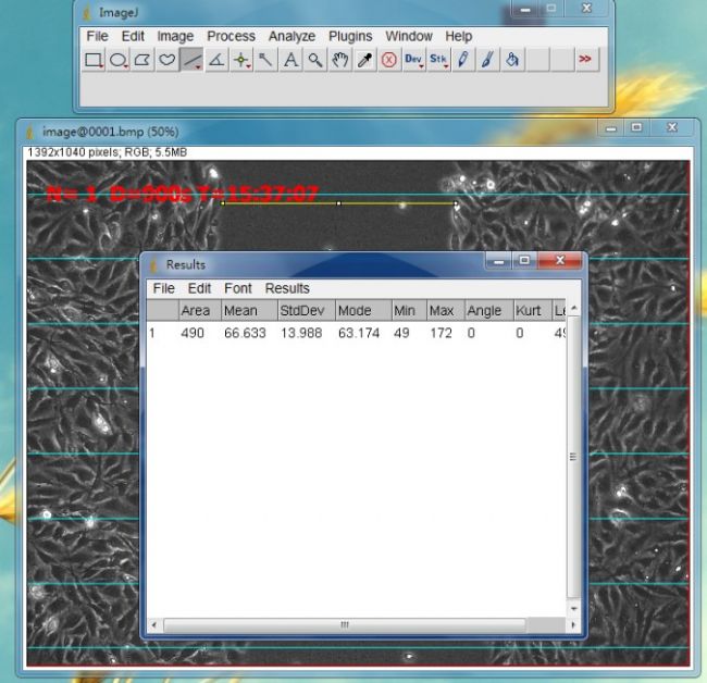
(6) Statistical method: After opening the picture using Image J software, randomly draw 6 to 8 horizontal lines to calculate the mean value of the distance between cells. 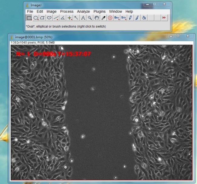

3. Note:
(1) When rinsing with PBS buffer, the adherent is slowly added to avoid the scattering of single-layer adherent cells, which affects the experimental photographing results.
(2) Generally, scratch tests are performed without serum or low serum (<2%), otherwise cell proliferation cannot be ignored.
(3) According to the vertical direction of the line drawn behind the 6-hole plate, a number of intersections can be formed as a fixed detection point to solve the problem that the position is not fixed when viewed from front to back.
two. Main problems of traditional experimental methods
The disadvantage of traditional experimental methods is that cell proliferation has an effect on the experimental results, even in the absence of serum or low serum conditions, the shape of the cells
State, metabolism, etc. will be affected, due to the overall down-regulation of the intracellular signaling system, the rate of cell migration will also
A lot slower; in addition, the creation of "wounds" may also be large and small, the edges are not neat and other errors;
It is almost impossible to achieve complete consistency at each observation point.
Â
three. The latest experimental solution ------- wound healing scratch plugin 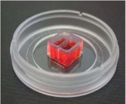
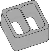
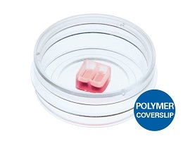
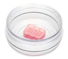
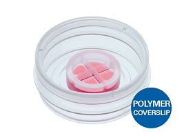
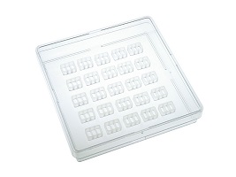
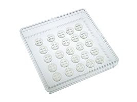
Adhesive bottom; biocompatible silicone material; 9 mm x 9 mm x 5 mm; two holes (70 μl/well); growth area per hole 0.22 cm2; produces a "gap" of 500 μm width   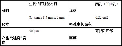
Features:
1. To some extent, the process of cell migration in vivo is simulated.
2. Ideal for studying cell migration with cell-to-extracellular matrix (ECM), cell-to-cell interactions.
3. Compatible with microscope systems including live cell imaging for analysis of cell-cell interactions.
4. The easiest way to study cell migration in vitro.
Â
Cell culture plug-in method compared with traditional scratch test
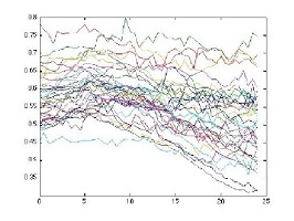
Cell culture plug-ins provide reproducible experimental results:
The left picture shows the experimental data statistics made by the wound healing plug-in; the right picture shows the experimental data statistics made by the head scratches. 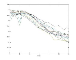
Summary of the program
( 1) The new cell scratch test method can ensure the standardization of scratches and ensure the repeatability of the experiment - compared with the scratches of the gun head, the scratches will be twisted and twisted, and it is impossible to guarantee the same every time;
(2) The new cell scratch test method can prevent the scratch of the gun head from injuring the coating; if the manual scratch marks the coating, it will directly affect the result of cell migration;
(3) Direct microscopic examination, the imaging effect is good;
(4) The new cell scratch test method has matching image analysis. The image analysis calculates the healing condition by calculating the blank area of ​​the experimental area, which is more objective and accurate than the analysis and calculation;
(5) Introduction to product usage: It is only necessary to plant cells in the two holes of the plug-in. When the cells are attached and then the plug-in is removed, a standard scratch will be left between the cells, and the cell healing can be started periodically. The situation
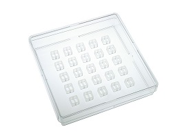
(6) A variety of specifications for different experimental procedures, 2 wells, 3 wells, 4 wells, with petri dishes or inserts, cell tracking experiments, special plug-in culture dishes, etc.